


What is osteochondrosis
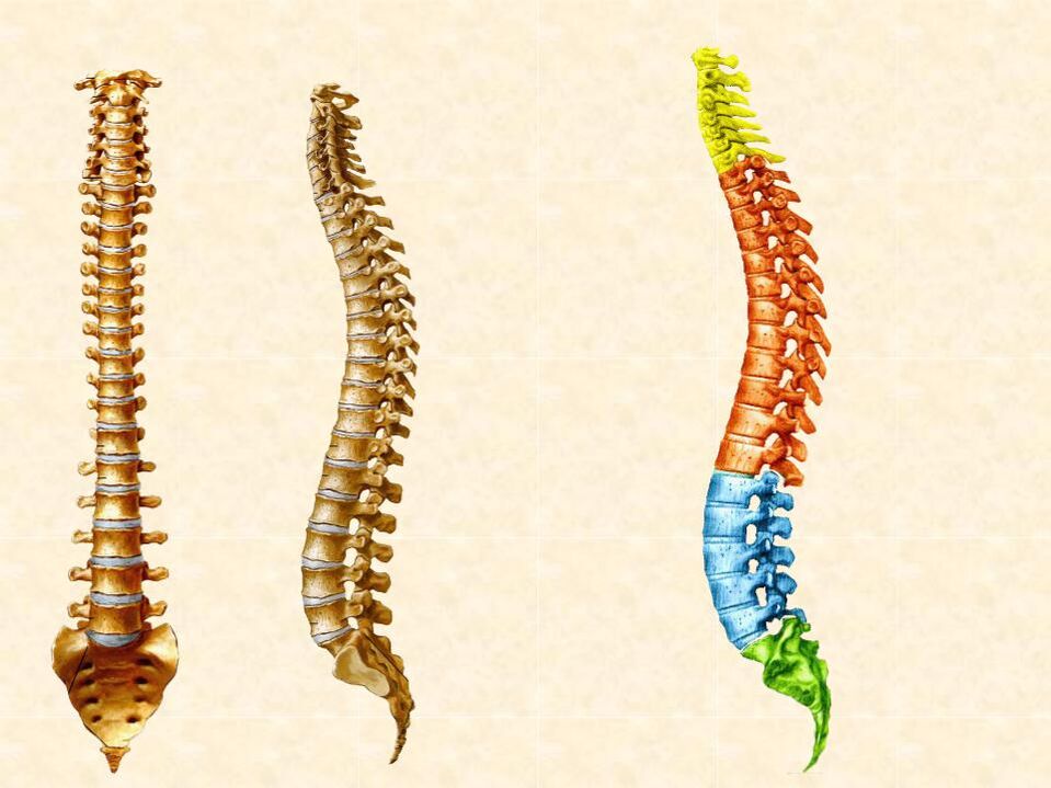
- Vertebrae are composed of vertebral bodies, vertebral arches, and vertebral processes. There are joints called facets between the arches of adjacent vertebrae
- Intervertebral discs are located between adjacent vertebrae
- spinal ligaments
- Posterior longitudinal, anterior longitudinal - passing along the front and rear vertebral bodies
- Ligamentum flavum - an arch that connects adjacent vertebrae
- Supraspinous and interspinous ligaments - connect the spinous processes
- The spinal cord is located within the spinal canal, from which nerve roots extend. They are nerve cell processes. Through these processes, the brain receives information about the state of the tissue and in response sends signals that regulate its function: muscle contractions, blood vessel diameter changes, and so on.
The intervertebral discs make up one-third of the length of the spine and act as shock absorbers, protecting the vertebrae from overloading when lifting heavy objects, standing or sitting for long periods of time, bending and twisting. Each disk contains:
- The nucleus pulposus, located in the inner center, contains large amounts of hyaluronic acid, type II collagen, which retains water. This gives the regular core a jelly-like consistency for effective cushioning. As degeneration progresses, the composition of the interior of the intervertebral disc changes, the water content decreases, the core "dries out", and the disc height decreases
- The annulus fibrosus is located outside the nucleus and is composed of 15-25 layers of collagen fibers. The collagen in the annulus fibrosus is type I. It is denser than the core and is needed to secure the inside of the disk and protect it from damage. The fibers of the ring are interwoven along the periphery with the posterior longitudinal ligament of the spine. This ensures the stability of the spinal structures in healthy people—a condition doctors call spinal stability. In people with degenerative diseases, the annulus fibrosus can rupture, so instability may occur: adjacent vertebrae may move forward or backward relative to each other. This is dangerous because the nerve roots between them are pinched
Stages of development of osteochondrosis
- Initial degradation.If the intervertebral disc does not receive adequate nutrition, it will wear out, lose height, and rupture. The nucleus pulposus protrudes through microlesions in the annulus fibrosus, irritating the posterior longitudinal ligament, causing back muscle pain and reflex spasms.
- Herniated disc.The fibers of the annulus fibrosus are destroyed, and the nucleus pulposus protrudes even more, forming a hernia. It can compress spinal nerve roots, causing paralysis or paralysis of limb muscles and decreased skin sensitivity. One of the complications of a hernia is its isolation—the separation of the herniated portion of the disc from its main portion.
- Progression of degeneration of the protrusions and other structures of the spine.The intervertebral discs become more compact and the body attempts to compensate for the excessive movement of the spine by forming pathological bone growth (osteophytes) in the vertebral bodies. Like the hernia itself, they can affect the nerves and ligaments, disrupting their function and causing pain. Unlike hernias, bone spurs do not dissolve.
Complications of osteochondrosis,In addition to compressing prominent spinal nerve roots:
- spondyloarthropathy. Reduced disc height puts more stress on the facet joints. They can develop inflammation and malnutrition, causing them to become "dry" and cause pain.
- SpondylolisthesisRelative displacement of the vertebral body due to ligament damage
- Degenerative processes in the ligamentum flavum area cause it to thicken. This is dangerous because the ligamentum flavum is adjacent to the spinal canal, narrowing the spinal canal and compressing the spinal cord.
- At the level of the 1st and 2nd lumbar vertebrae, extending downward from the spinal cord"ponytail" - A bundle of nerve roots responsible for the innervation of the lower limbs and pelvic organs: bladder, rectum, external genitalia. Cauda equina syndrome is one of the most dangerous complications of osteochondrosis, manifested by severe pain, leg muscle weakness, and perineal numbness, incontinence.
Causes of Osteochondrosis of the Back
It's impossible to accurately predict how severe a given person's degenerative changes will be and whether they will lead to complications. There is a genetic predisposition to osteochondrosis, but the specific genetic mutations responsible for the course of the disease have not been identified. Therefore, there is no accurate genetic test that can show an individual's risk. There are certain factors that increase the risk of osteochondrosis. They are targeted for osteochondrosis preventive measures.
Risk factors for osteochondrosis include:
- Excessive load on the spine: Professional sports, weightlifting, frequent heavy physical labor
- Staying still or in incorrect posture for a long time:Sitting, slouching, crossing legs, sitting in a chair without lumbar support, working in an inclined vertical position
- sedentary lifestyleResulting in weakness of trunk muscles and inability to effectively support the spine
- overweight1. Obesity puts extra pressure on your back and joints
- Smoking- Nicotine and other ingredients in cigarettes disrupt the diffusion of nutrients from blood vessels to tissues, including the spinal discs
- alcohol intake- Regular consumption can lead to malabsorption of calcium from food. Calcium deficiency leads to reduced vertebrae density
- Back injuryThe vertebrae or disc structures are damaged so the recovery process is much slower than the degeneration process
Spinal Osteochondrosis in Adults: Symptoms
- Degeneration of the cervical spine can cause muscle stiffness, neck pain that radiates to the shoulders, arms, or back of the head, and worsens with head movement
- Changes in the thoracic spine rarely occur because it is the most static. If a hernia does occur, pain will occur between the shoulder blades
- Lumbar hernias are more common than hernias elsewhere and present as pain in the lower back or sacrum that radiates to the buttock area and legs. Stiffness in the lower back was also noted. The pain worsens when you sit, stand, and bend for long periods of time.
- Muscle weakness in the limbs until paralysis
- Skin sensitivity disorder of extremities
- Bladder and rectal dysfunction with lumbar radiculopathy
Spinal osteochondrosis in youth
Spinal Osteochondrosis: Treatment
In many cases, uncomplicated hernias will heal on their own. The resorption process may be accompanied by the formation of excess connective tissue and calcification in the spine, which increases the likelihood of future recurrence of the disease. Available physical therapy techniques and special exercises can help:
- Accelerate the absorption of hernia
- Increase disk power
- Normalize biomechanics of movement and load distribution
- Avoid the need for surgery in the future
- MLS Laser Therapy - The laser radiation used has anti-inflammatory effects, dilates lymphatic vessels, and improves lymphatic drainage
- Acupuncture - This method relieves pain, swelling and inflammation caused by the body's reflex response to special needles stimulating biologically active points in the body
- Magnetic therapy stimulates blood flow, normalizes the diffusion of nutrients, and removes toxins from the thickness of the disc, accelerating the recovery process
- Therapeutic Physical Education - Special sets of exercises help strengthen trunk muscles, learn how to properly distribute load on the back, maintain correct posture, and relieve muscle spasms. To monitor performance, it is best to start working with a coach and then continue practicing on your own as recommended